Diagram Of Hip.and Back.muscles : Pin On On Being Human / Deadlift muscles will include knee, hip, and back extensors, which primarily include the quads, glutes, and spinal erectors.
Diagram Of Hip.and Back.muscles : Pin On On Being Human / Deadlift muscles will include knee, hip, and back extensors, which primarily include the quads, glutes, and spinal erectors.. Human muscle system, the muscles of the human body that work the skeletal system, that are under voluntary control, and that are concerned with movement, posture, and balance. Some of these muscles are quite large and cover broad areas. The back's muscles start at the top of the back (named the cervical vertebrae) and go to the tailbone (also named the coccyx). Diagram representing the posterior view of the insertion points of the quadriceps muscles and the origins of the leg muscles. They are attached to the femur (thighbone), tibia (shinbone), and fibula (calf bone) by fibrous tissues called ligaments.
Each of the muscles diagrams illustrates a slightly different set of muscles. Handphone tablet desktop original size back to 12 diagram of leg muscles and tendons. Muscles/tendons flashcards from molly m. The hip muscles are all the muscles that act on the hip joint. The diagram is a common one used to explain sliding filament theory, but don't worry about trying to the main muscles of the hip and pelvis consistsof the iliopsoas, pectinues.
Muscles Of The Hips And Thighs Human Anatomy And Physiology Lab Bsb 141 from s3-us-west-2.amazonaws.com The muscular system consists of various types of muscle that each play a crucial role in the function of the body. Nine may seem like quite a lot, but these muscles are essential for creating the wide range of hip movements used by dancers, sportspeople and music lovers. Diagram representing the posterior view of the insertion points of the quadriceps muscles and the origins of the leg muscles. Back muscles anatomy lower back muscles anatomy human anatomy. Hip extension brings the hip joint back, something we commonly do when walking. Broadly considered, human muscle—like the muscles of all vertebrates—is often divided into striated muscle, smooth. The back comprises the dorsal part of the neck and the torso (dorsal body cavity) from the occipital bone to the top of the tailbone. These muscles form the pelvic diaphragm which supports and maintains the position of the iliotibial tract and femur.
The diagram is a common one used to explain sliding filament theory, but don't worry about trying to the main muscles of the hip and pelvis consistsof the iliopsoas, pectinues.
Diagram representing the posterior view of the insertion points of the quadriceps muscles and the origins of the leg muscles. While the thigh muscles will be slip into the anterior, medial and posterior groups. This is a table of skeletal muscles of the human anatomy. Common hip and back pain causes include injury to muscles from overuse disc injurydegeneration or spinal stenosis. In human anatomy, the muscles of the hip joint are those muscles that cause movement in the hip. It joins the lower limb to the pelvic girdle. Prime movers cross hip joint anteriorly: Required to throw a baseball, swing a bat or golf club. Learn with flashcards, games and more — for free. Tendons attach the muscles to each other. The back comprises the dorsal part of the neck and the torso (dorsal body cavity) from the occipital bone to the top of the tailbone. Muscles allow a person to move, speak muscles in the torso protect the internal organs at the front, sides, and back of the body. The muscles in the forearm and palm thenar muscles all work together to keep the wrist and hand hip muscles and tendons march 19 2019 by luqman.
Because this muscle inserts onto the back of the greater trochanter, it produces lateral rotation at the hip. Here we will look at the gluteal muscles and the inner hip muscles. The back's muscles start at the top of the back (named the cervical vertebrae) and go to the tailbone (also named the coccyx). To learn more about the lower back anatomy of the spine, please watch this video. There are anterior muscles diagrams and posterior muscles diagrams.
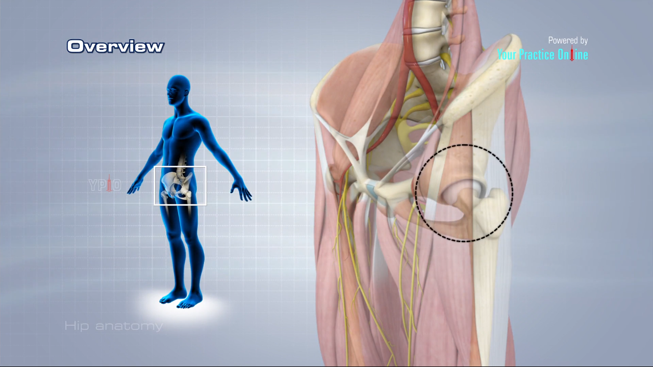
Hip Anatomy Video Hip Orthopaedics Videos Your Practice Online Education from www.ypo.education Luckily you've found this page to help you. Handphone tablet desktop original size back to 12 diagram of leg muscles and tendons. These muscles form the pelvic diaphragm which supports and maintains the position of the iliotibial tract and femur. Here we will look at the gluteal muscles and the inner hip muscles. The back comprises the dorsal part of the neck and the torso (dorsal body cavity) from the occipital bone to the top of the tailbone. Insertion because that just makes it more confusing and your muscles don't really identify themselves that way anyhow… Extension and lateral rotation at the hip. Related posts of muscles of the lower back and hip diagram muscle anatomy posterior.
This is a diagram of the larger and more surface muscles of the low back.
Nine may seem like quite a lot, but these muscles are essential for creating the wide range of hip movements used by dancers, sportspeople and music lovers. Learn with flashcards, games and more — for free. It is opposite from the chest, and the vertebral column runs down. Abduction and medial rotation at the hip. Luckily you've found this page to help you. Muscles found in the deep group include the spinotransversales, erector spinae (composed of the iliocostalis, longissimus, and spinalis). Deadlift muscles will include knee, hip, and back extensors, which primarily include the quads, glutes, and spinal erectors. Extension and lateral rotation at the hip. The hip muscles are all the muscles that act on the hip joint. Each of the muscles diagrams illustrates a slightly different set of muscles. There are anterior muscles diagrams and posterior muscles diagrams. The bones of the spine and the ribs provide further protection. Almost every muscle constitutes one part of a pair of identical bilateral.
Tendons attach the muscles to each other. Abduction and medial rotation at the hip. Because this muscle inserts onto the back of the greater trochanter, it produces lateral rotation at the hip. Globular end of the femoral neck. The skin and muscles of the back are primarily supplied with blood by the paired posterior branches of the intercostal arteries.
Lower Back Pain And It Band Stretching Belymbr from 143kmn1cemus16arfe3f8cly-wpengine.netdna-ssl.com Prime movers cross hip joint anteriorly: Hip flexor muscles and attachments. Most modern anatomists define 17 of these muscles, although some additional muscles may sometimes be considered. The muscles that affect the knee's movement run along the thigh and calf. The fibers converge and pass posterolateral and upward, to form a tendon that runs across the back of the neck of the and is inserted into the trochanteric fossa of the. Here we explain the major skeletal muscles, muscle structure, fibre types, contractions and sliding filament theory. This article considers the hip joint specifically. • the sciatic nerve passes just inferior to the piriformis therefore a tight piriformis muscle my contribute to compression on the sciatic nerve.
Abduction and medial rotation at the hip.
Here we explain the major skeletal muscles, muscle structure, fibre types, contractions and sliding filament theory. Learn with flashcards, games and more — for free. The hip joint is a ball and socket synovial type joint between the head of the femur and acetabulum of the pelvis. Back muscles anatomy lower back muscles anatomy human anatomy. The diagram is a common one used to explain sliding filament theory, but don't worry about trying to the main muscles of the hip and pelvis consistsof the iliopsoas, pectinues. Here we will look at the gluteal muscles and the inner hip muscles. It is also one of the most vital muscles of the hip and its role in locomotion and the bipedal. Most modern anatomists define 17 of these muscles, although some additional muscles may sometimes be considered. It is opposite from the chest, and the vertebral column runs down. Dislocation of the hip joint. This article considers the hip joint specifically. This is a table of skeletal muscles of the human anatomy. • the sciatic nerve passes just inferior to the piriformis therefore a tight piriformis muscle my contribute to compression on the sciatic nerve.


0 Komentar